
Transport in mammals (complex multicellular organisms) is carried out by a circulatory system of arteries, veins and capillaries.
This is because all organisms require food, water, oxygen and other minerals for growth, development and repair of worn-out parts of the body.
Similarly, an efficient system is required to remove waste products from the body. The circulatory and excretory system (collectively) carry out transport in animals.
This was all about the introduction of this topic. Now, let’s discuss some other important concepts without further introductions.
Transport in Mammals
Single Circulation in Fish:
You might be wondering, why is it important to know about the single circulation in a fish?
Fish are not as active as other animals and their single circulatory system is enough to fulfil their needs. However, more active animals, such as mammals, need double circulation to fulfil their needs.
We will take a look at double circulation in mammals in detail later on in this article.
There is a single systemic circuit for blood (in fishes). The heart pumps the blood to the gills (respiratory organ found in many aquatic organisms) to become oxygenated and then, the blood flows to the rest of the body and then back to the heart.
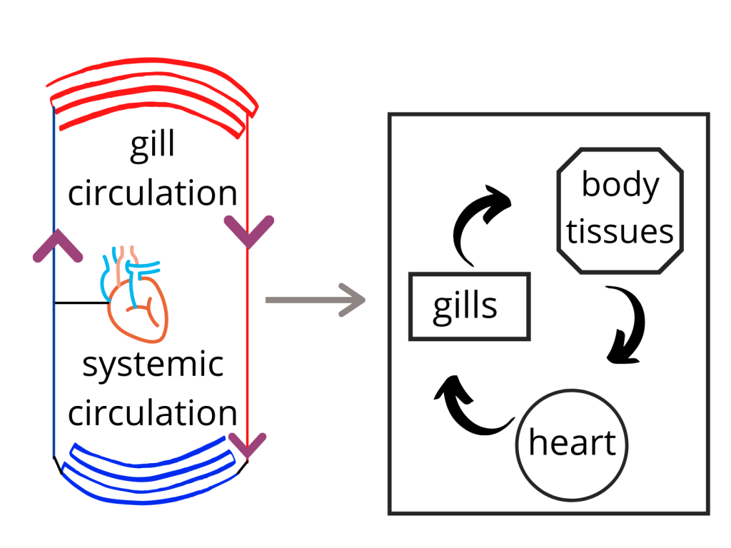
The two-chambered heart of the fish has a single atrium and a single ventricle. The atrium collects the blood from the body and then sends it to the ventricle which pumps it to the gills.
This movement of blood is also referred to as systemic circulation (movement of blood between the heart and gills).
When talking about blood, what comes to your mind? Let’s discuss that.
Blood:
You would have definitely heard of this term but we need to look at it from an examination point of view.
Blood is also known as fluid tissue or tissue fluid, but what is the reason for that?
This is because the blood contains Plasma, Red blood cells and White blood cells. It is a tissue because it is a collection of (similar) specialized cells that are suspended in a liquid Plasma.
With that being said, I have a question for you over here. Is tissue fluid another name for blood?
When we talk about Plasma, it is a yellowish liquid that comprises mostly of water and some other constituents are:
- Food substances (glucose and amino acids).
- Mineral salts.
- Soluble proteins.
- Hormones and (excretory) products such as Urea and Uric acid.
As discussed above, red and white blood cells are also major components of blood. So, let’s take a look at them in detail now.
Red blood cells (RBC’s):
There are multiple ways of referring to this blood component because these cells are also known as erythrocytes and red blood corpuscles.
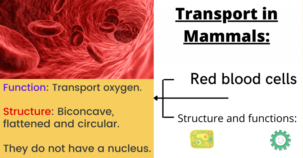
Their primary function is to transport oxygen to the body tissues through the blood flow (circulatory system). These cells are biconcave, circular and flattened (in shape) and they do not contain a nucleus.
This is because it ensures that they can carry more haemoglobin (a red iron-rich protein inside red blood cells) and ultimately, more oxygen is transported.
These cells are made in the bone marrow and their average life-span is 120 days and after that, they are replaced with new cells.
The function of these cells is to carry oxygen from our lungs to the rest of the body and when they return back to the lungs, the bring carbon dioxide to be exhaled.
Although red blood cells are considered cells, yet they lack organelles such as a nucleus, DNA and mitochondria (double membrane-bound organelle to generate energy).
Actually, these cells not having a nucleus is actually an adaptation so that they are better equipped for their task (carrying more oxygen).
This is some information that you should be aware of. Now, it is time to discuss the structure and function of white blood cells.
White blood cells:
White blood cells (also known as leukocytes) are the cells of the immune system (system to protect the body from infections and diseases).
In other words, these cells help your body fight infections and they are much fewer than the red blood cells (composing approximately one per cent of your blood).
Some of the functions of these cells are:
- Phagocytosis
- Formation of antibodies
- Tissue rejection
Unlike red blood cells, white blood cells are larger in size and they have a nucleus and mitochondria. The life-span of these cells ranges from 13 to 20 days after they are released from the bone marrow.
To understand the function of white blood cells, you need to know the process of phagocytosis.
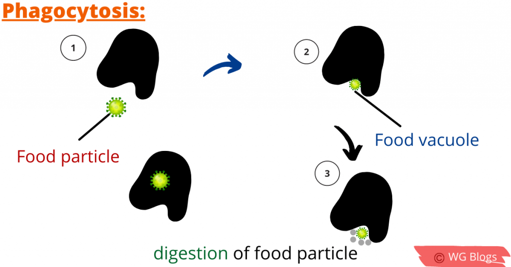
Phagocytosis is a process in which the phagocytes (living cells) engulf other particles or cells. This is a defensive reaction by the body against infection and antigens (foreign substances).
Dead tissue cells, bacteria, dust particles, pigments and other foreign bodies are commonly phagocytosed by the white blood cells.
The size of the particle plays an important role in the speed at which the phagocytic cell engulfs the particle. For example, larger objects such as lumps of bacteria take a longer time to be engulfed.
Further reading:
Excretion | GCE O level Biology Notes
Energy changes | GCE O level Notes
The pathogen is broken down in order to destroy it and in this way, the white blood cells get rid of the harmful materials for the body.
Note: The white blood cells are colourless.
Now, what is tissue rejection?
Let’s suppose that both kidneys of a person fail to work and they take a kidney from another person. The cells in the new kidney will be recognized as “foreign” by the immune system.
As a result, the body will attack and destroy them and since the immune system is triggered, antibodies are secreted and the organ may be rejected. This is some basic information about tissue rejection.
The reason being that white blood cells recognize the body tissues (as there is a set of antigen on the surface of each cell).
We also discussed that platelets are a major component of the blood as well. So, let’s discuss some important concepts regarding platelets as well.
What are platelets?
These small and colourless cell fragments in our blood are also known as thrombocytes and their primary function is clotting of blood to stop or prevent bleeding.
Thrombocytes (platelets) are produced in our bone marrow and they are essential in surviving surgeries during organ transplants and other chronic diseases.
You should also know that the number of platelets in our body can reduce significantly due to the following reasons:
- Excessive bleeding.
- When a person’s bone marrow is damaged.
- There might be some infections or medicines which have destroyed the platelets.
Now, how does clotting occur? Let me tell you this process, step-by-step.
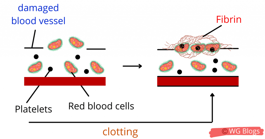
Firstly, the damaged blood vessel will cause thrombokinase (an enzyme) to be secreted. This enzyme will convert prothrombin (a protein) to thrombin.
The thrombin will cause the conversion of soluble protein fibrinogen to insoluble fibrin threads and in this way, clotting stops the excessive loss of blood.
Now, it is time to move learn about the circulatory system in mammals and find out how transport in mammals actually takes place.
The circulatory system in animals:
The blood circulation in our body is ensured by a muscular pump, the heart, and the valves which prevent the backward flow of the blood so that blood moves in only one direction.
The circulatory system is the composition (network) of the blood vessels – the arteries, veins and capillaries. When the heart relaxes, the blood is filled while when it contracts, the blood moves out and moves through the blood vessels.
Arteries, Veins and Capillaries:
The arteries take blood away from the heart and provide oxygen-rich blood (except the pulmonary artery) to the tissues of the body.
The arteries are elastic, thick and muscular structures have smooth tissues because the blood flows through them with great pressure and these capabilities help them to support it.
As a result, the artery is capable of stretching and coming back to its normal position which ensures the flow of blood.
Note: The largest artery in our body is the aorta that supplies the blood from the left ventricle to the remaining body. We will look at the structure of the heart later in this article.
The pulse you feel is due to the contraction and expansion of the arteriole wall due to the beating of the heart and the flow of blood with high pressure.
- When arteries dilate, their lumen becomes wider (more blood flows).
- When arteries contract, their lumen becomes narrower (fewer blood flows).
Veins are the blood vessels that carry blood towards the heart. They mostly carry deoxygenated blood (except the pulmonary vein).
The blood pressure in veins is lower as compared to arteries and therefore, they are not as muscular and thick (comparatively). Moreover, the smallest veins in the blood are called venules.
However, Vena Cava is the largest vein in the body.
The most important point over here is that veins have valves (the structures which prevent the backward flow of blood.
Why do veins need valves?
This is because the blood flows through them with low pressure and there is a chance of the blood moving backwards which is prevented by the valves.
This ensures that the blood flows in only one direction. Cool, isn’t it?
Capillaries are small vessels that branch from the arterioles and these thin vessels allow the exchange of material between the cells.
Simply, they carry blood from the arteriole (small artery) to the venule (small vein) and allow the exchange of gases, nutrients and waste between the blood and tissue.
Now when you know about these blood vessels, it is time to discuss how the blood flows in our entire body.
Blood circulation in animals:
- Firstly, the right atrium (one of the four chambers of the heart) receives deoxygenated blood from the Vena Cava.
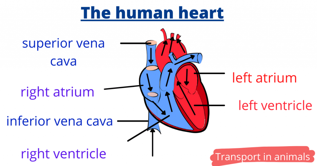
The upper (superior) vena cava brings blood from the upper parts of the body such as arms, head and neck while the inferior vena cava brings blood from the lower parts of the body such as the legs.
- Then, the right atrium contracts which cause the blood to flow to the right ventricle and the tricuspid valve opens.
This valve separates the upper chamber (right atrium) from the lower chamber (right ventricle) and the right and left part of the heart are separated by a muscular wall – Septum.
- When the right ventricle contracts, the blood moves through the pulmonary arteries to the lungs.
In the lungs, the blood becomes oxygenated through the exchange of materials (such as oxygen enters the blood and waste products leave the blood).
- Then, the blood returns back to the left atrium of the heart from the lungs via the pulmonary vein (carrying oxygenated blood).
When the left atrium contracts, the bicuspid valve opens so that the blood moves to the left ventricle. When the left ventricle contracts, the blood is pumped to the entire body (through the aorta).
This is how to double circulation takes place in mammals and allow the entire body to have a constant supply of blood.
The concept of diastole and systole:
These two terms are related to the contraction and relaxation of the heart muscles.
Simply, the period of contraction during the blood flow is systole while the period of relaxation is diastole. Of course, this is not so convincing and let me explain it further.
Note that both atriums (left and right) of the heart contract and relax simultaneously (at the same time). Similarly, both ventricles contract and relax at the same time.
When the ventricles contract, the tricuspid and bicuspid valves close suddenly producing a “lub” sound – systole (during which blood pressure increases).
When the ventricles relax, the semilunar valves close to prevent the backflow of blood-producing a “dub” sound – diastole.
I know this may be a bit confusing to you but just remember that ventricle contraction refers to systole while ventricle relaxation is diastole.
The systole and diastole makes up one heart beat.
Coronary heart diseases:
The heart muscles receive blood from the coronary arteries (just like the renal artery to the kidney).
If these arteries are blocked due to fatty deposits (called “plaques”) containing cholesterol, coronary heart diseases may occur. This is because the supply of blood is disturbed and if the heart fails to contract, one may face a heart attack.
Some of the possible causes of coronary heart diseases are:
- Stress and anxiety increase blood pressure.
- The blood vessels may constrict due to smoking (rise in blood pressure).
- Poor diet consumption can increase cholesterol levels if the diet contains saturated fats (in excess).
- Males and people of higher age are more likely to develop coronary heart diseases.
The blockage of these blood vessels can be caused by arteriosclerosis (walls of arteries become thick and hard).
In order to prevent these diseases, you should take a healthy diet (reduce animal fats) and consume more vegetable and fruits. Moreover, regular exercise regulates blood pressure as well.
If one quits smoking, the risk of these heart diseases can fall drastically as well because the damage of arterial walls can be prevented.
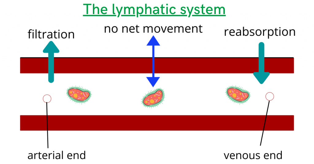
The lymphatic system:
Since the walls of capillaries are thin, the substances such as water, mineral salts and dissolved gases can leak out because of the blood pressure.
It is to note that the white blood cells can also leak out during this process and as a result, this blood plasma gives rise to lymph fluid.
But, let me get into more detail and provide a greater insight of the topic.
This portion of blood that escapes from the blood capillaries is known as interstitial fluid or extracellular fluid and contains glucose, oxygen, amino acids and some other nutrients as well.
However, the majority of this fluid seeps back into the bloodstream.
This system also plays a vital role in defending the body against infections (by producing white blood cells). The vessels that collect the lymph fluid and return it back to the blood are known as lymphatic vessels.
So, we can say that the purpose of the lymphatic system is to return the tissue fluid (plasma without proteins) back to the blood.
Now, I have a question for you over here. Why does this fluid does not contain protein and red blood cells?
The answer is that they are simply larger and cannot pass out through the capillary tube.
Conclusion:
With this, our topic about transport in mammals has come to an end.
Now, it is your turn.
What part of this topic do you find the most interesting? I hope you will know about important concepts such as double circulation in animals, coronary heart diseases and the lymphatic system now.
But before concluding, I have a question for you over here? How does the transfer of material between the capillaries and the tissue fluid take place? Try to find out the answer to this question from the above information.
Thank you very much for reading and staying with me till the end. Stay tuned for more.