
Do you know that a human eye (organ of the visual system) blinks about 4,200,000 times a year (on average)?
Amazing, isn’t it?
You should also know that the eyes are responsible for vision in animals. They detect light and form images. But, do you about the internal structure of the eyes?
Do not worry! I will explain all these topics to you in detail. So, let’s dive into the topic without further introductions.
Sense Organs:
Sense organs are specialised organs that allow us to respond to the changes in our environment.
But, why do humans need sense organs.
Simply, to survive. This is because sense organs allow us to make adjustments to the changes in the environment. So basically, you can take it as “negative feedback” as we discussed in the topic of homeostasis.
Eye is a sense organ and there are five sense organs in total. These are:
- Eyes
- Ears
- Nose
- Tongue
- Skin
I like to refer to these organs as “neural networks“. Do you know why?
This is because these organs are composed of sensory neurones (nerve cells that transmit nerve impulses from the sense organs to the central nervous system).
This is the reason why these organs help us to respond to the changes in the environment (in order to survive).
Now, let’s take a look at the structure of the eyes in detail.
Structure of the Eye:
(Front view):
Before discussing the internal structure of the mammalian eye, let’s take a look at the front view of an eye.
Eyes are located in the bony cavities (which are known as the orbits) of the skull. Their movement is controlled by six extraocular muscles (rectus muscles).
First of all, you should know that a thin layer of conjunctiva covers the exposed part of the eye. This transparent mucous membrane helps to lubricate the eye (by producing tears and mucus).
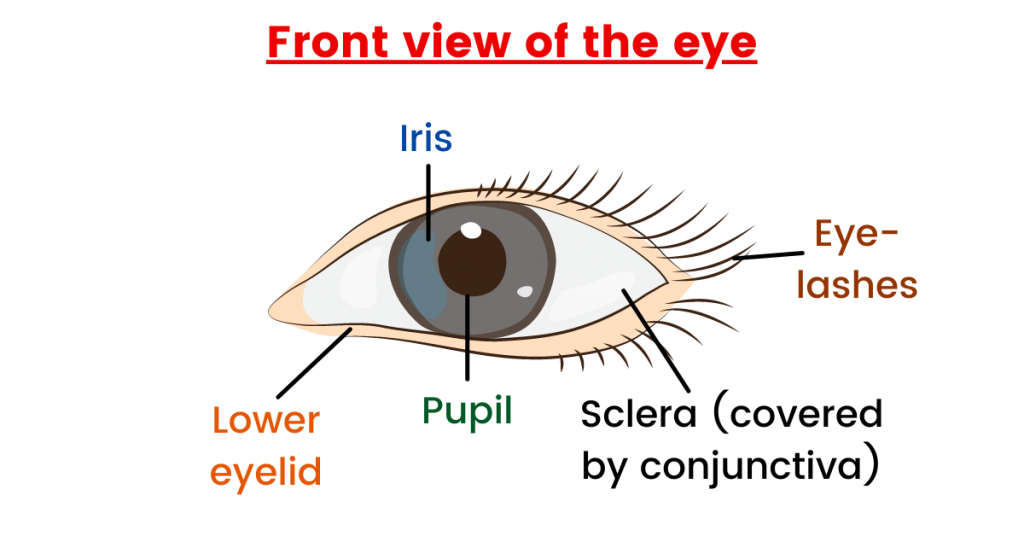
In the diagram above, you can also see eye lashes. But, what is their function?
The main function of eyelashes is to protect your eye tissues from dirt, dust and other harmful particles. So, they act as a barrier for the defence of the eye against harmful substances.
Moreover, we have eyelids (movable tissue) as well. These are the folds of the skin that close to protect our eye. In simple words, I explain them as a shield that protects the eyeball from injury or damage.
Eyelids also help us to spread tears (to lubricate the eye) which keeps the surface moist.
Summary:
- Conjunctiva: Lubricates the eye.
- Eyelashes: Protect the eye from foreign bodies (such as dust and dirt).
- Eyelids: Protect the eye and keeps the surface moist.
Now, let me take you through the internal structure of the human eye.
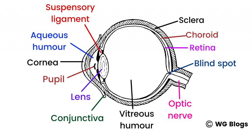
(Vertical section of the human eye):
If you view the human eye vertically, you will notice three layers (that make up its wall). These layers are:
- Sclera (outermost layer)
- Choroid (the middle layer)
- Retina (Inner layer)
The sclera (also known as the sclerotic coat) is a white layer that covers most of the outside of the eyeball. This fibrous and protective outer layer is in contact with the cornea (at the front).
Then comes the choroid coat. You should know that the choroid is a vascular layer and has connective tissues. Similarly, its function is to maintain the temperature and volume of the eye. It also provides nutrients to the outer retina.
Choroid is pigmented black to prevent the internal reflection of the light.
When we talk about the retina, it is a thin layer of tissue (and is the innermost layer of the eyeball). From the diagram, it is visible that it is located near the optic nerve.
The main function of the retina is to provide vision to mammals. But, do you how it happens? Let me tell you all this, step-by-step.
- Firstly, the retina receives the light that was focused on by the lens.
- Then, it converts the light to neural signals (which are sent to the brain).
- This allows image formation which you can also call “visual recognition”.
When we are talking about the retina, we have to talk about photoreceptors (these are the cells in the retina that respond to light).
The two kind of photoreceptors are:
- Rods (they are responsible for vision at low light).
- Cones (they are responsible for colour vision – Simply, cones allow you to see the light of different colours).
Summary:
- Sclera: For protection against mechanical injury.
- Choroid coat: Prevents the internal reflection of light (because it is pigmented black).
- Retina: Responsible for sensing light and colours.
Moving on, let me tell you about other parts of the human eye such as iris and the pupil.
Iris is a thin structure that is responsible for controlling the diameter (size) of the eye. The colour of your eye is the colour of your iris.
Further reading:
Transport in Plants Made Simple | GCE O Level
Transport in Mammals | Detailed Notes
Excretion explained | GCE O Level Biology Notes
So simply, you can say that the iris is responsible for the amount of light reaching the retina (we will take a look at this concept in detail, later in this article).
When we talk about the pupil, it is a hole that is located at the centre of the eye. This structure of the eye limits the light reaching the retina.
For example, during dim light, the size of the pupil increases to allow more light to reach the retina. However, in bright light, the size of the pupil decreases to allow less light to reach the retina.
With that being said, let me introduce you to the lens. This structure is located behind the iris and the pupil.
The lens is elastic, which means that it can change its shape (to create a clear image of the objects). So, it helps the light to be focused on the back of the eye (retina) for image formation.
Note: The lens are made up of elongated cells and they have no blood supply! But, how do they get their nutrients? For the answer, you have to study the surrounding fluids – aqueous humour and vitreous humour.
If you clearly look at the lens in the diagram, you will notice that it divides the eye into two chambers. The chamber at the front (the smaller one) is filled with aqueous humour.
Aqueous humour is a transparent water-like fluid (slightly alkaline liquid). And for your knowledge, let me tell you that it is similar to the human’s blood plasma (except that it contains less glucose and proteins).
So, the answer to the above question is that this fluid (aqueous humour) provides nutrients to the lens. In short, the aqueous humour is fluid in the eye (which maintains the eye in a pressurized state).
Moving on, let me introduce you to vitreous humour.
It is a jelly-like fluid that occupies the space between the lens and the retina of the eyeball. If I talk about its function, it is responsible to maintain the shape of the eye.
Moreover, it also acts as a “shock absorber” (because it absorbs shocks to the eye).
Both aqueous and vitreous humour refract light and keep the eyeball firm.
Summary:
- Iris: It controls the amount of light that enters the eye.
- Pupil: It is the path for light (allows light to enter the eye).
- Lens: It allows light to be focused on the retina.
- Aqueous and vitreous humour: They keep eyeball firm and refract light.
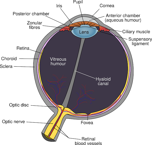
Now, let me introduce you to fovea (yellow spot) and the blind spot.
The fovea is a spot on the retina where the vision is the sharpest (clearest). This means that this is the place where images are normally focused.
Note: This is the place where the concentration of cones (related to colour vision) is the highest (because light falls directly on them at this spot).
When it comes to the blind spot, you should know that it is the region where there are no photoreceptor cells (rods and cones). So, what does this means?
This means that no image formation takes place in this area. Therefore, it is known as the blind spot because you cannot see anything over here. Now, let’s discuss some other important concepts in this topic as well.
The role of circular and radial muscles:
As I explained earlier, our eye takes different actions to adjust to the changes in the environment. For example, our muscles perform differently in bright and dim light.
But, what is the difference? Let me tell you all this in detail.

In bright light, our pupil becomes smaller in size to allow less light to enter the eye. This is because we do not want too much light to enter our eyes.
So in bright light, our eyes takes the following actions:
- Circular muscles contract.
- Radial muscles relax.
- The pupil constricts (becomes smaller).
On the other hand, can you guess what happens in dim light? Yes, you are right. The exact opposite happens.
In dim light, our eyes takes the following actions:
- Circular muscles relax.
- Radial muscles contract.
- The pupil enlarges (becomes bigger in size as compared to normal).
The circular and radial muscles are two involuntary muscles in the iris. This means that when one contracts, the other relax (antagonistic muscles) and vice versa.
A thing to note over here is that the circular muscles are like a circle (so they are around the pupil). Similarly, radial muscles (as the name suggests) are like a radius because they connect the pupil to the iris.
Pretty simple, isn’t it?
Now, let’s take a look at some other topics such as vision and focusing (also called accommodation).
Vision and Focusing:
Before diving into the topic, let me introduce you to some structures in the eye. These structures allow us to carry out focusing (accommodation).
Suspensory ligaments: These are the series of fibres that connect the lens with the ciliary body of the eye.
Ciliary muscles: These elongated muscles (longitudinal and circular fibres) change the shape of the lens. These muscles occupy the largest space in the ciliary body.
I introduced these structures to you because they allow us to form clear images. You should also know that the image formed on our retina is inverted (upside down), diminished (smaller in size) and reversed.
But, our brain corrects this and therefore, we are able to see properly.
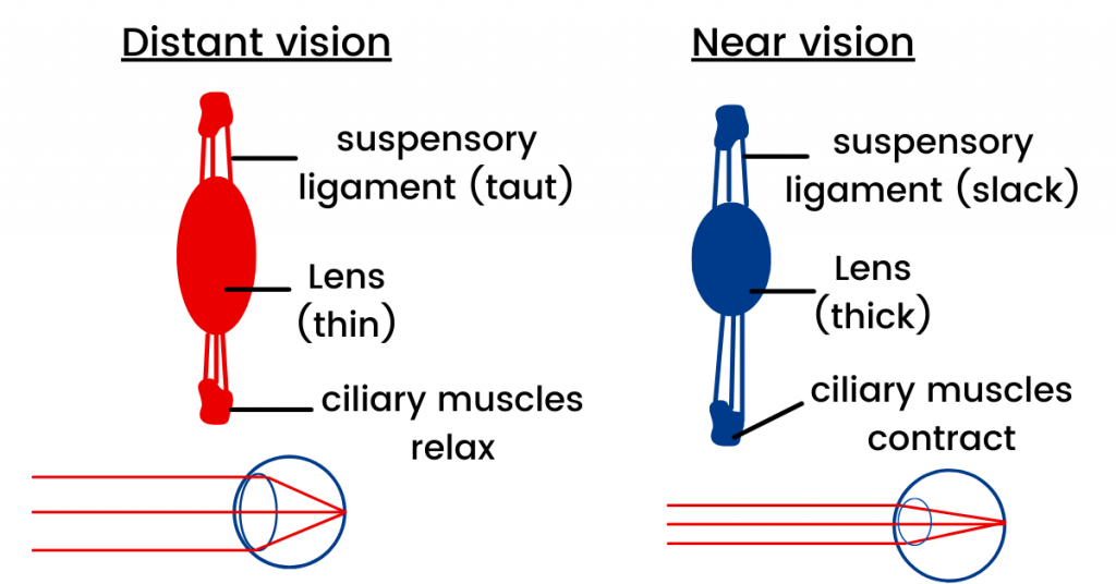
Focusing for near vision:
When you look at a near object, the following things happen:
- Circular muscles of the ciliary body contract (shorten).
- The suspensory ligaments slacken (loosen).
- The lens becomes more rounded (thicker).
Due to all this, clear image forms on the retina. This is known as accommodation. You should also know that the nearer the object, the more circular muscles contract (so the lens become more convex). But, there is a limit to it as well.
Moving on, let’s discuss what happens if we have to focus for distant (faraway vision).
Focusing for distant (far) vision:
If you look at a distant object, the circular muscles of the ciliary body relax. Moreover, the suspensory ligaments tighten (stretch).
Due to this, the lens comes in a flattened shape. This allows us to see the distant (far) objects clearly. To summarise, the following things happen when you look at a faraway object:
- The ciliary body relax.
- The suspensory ligaments become taut.
- The lens becomes less convex (flat).
Conclusion:
With this, our topic about the structure of the eye has come to an end. I have done my work. Now, it is on you.
I recommend you solve plenty of past paper questions to ace this topic. The topics covered in this article are animal receptor organs, eye structure, focusing and accommodation (under the topic of coordination and response).
Thank you very much for reading and staying with me till the end. Stay tuned for more.